
Contents
I. Introduction

Welcome to “Understanding Your Dog’s Body: A Detailed Anatomy Overview.” In this article, we will delve into the fascinating world of canine anatomy, exploring the various systems and structures that make up your furry friend’s body. Whether you’re a dog owner, a veterinary professional, or simply curious about the inner workings of man’s best friend, this article aims to provide you with a comprehensive understanding of your dog’s anatomy.
Throughout this article, we will break down the different body systems, including the skeletal, muscular, circulatory, respiratory, digestive, and nervous systems. We will explore the unique characteristics and functions of each system, shedding light on how they contribute to your dog’s overall health and well-being.
By gaining a deeper understanding of your dog’s anatomy, you will be better equipped to recognize signs of illness or injury, make informed decisions about their care, and provide them with the love and attention they deserve.
So, let’s embark on this journey together and explore the intricate and awe-inspiring world of your dog’s body. Get ready to uncover the mysteries that lie beneath that wagging tail and those adorable floppy ears!
II. The Skeletal System of a Dog

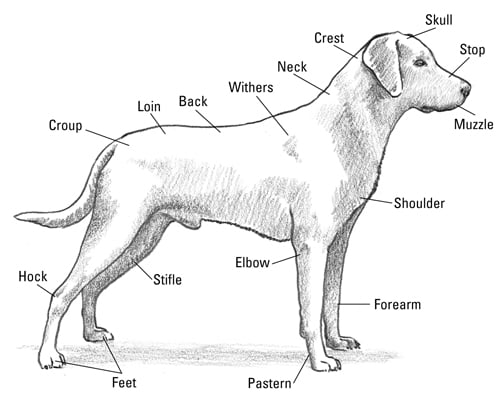
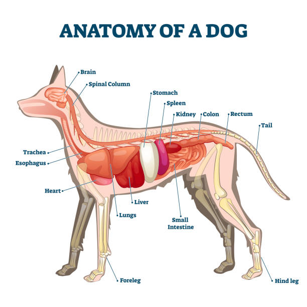
In this section, we will explore the skeletal system of a dog, focusing on the bones of the skull, spine, and limbs. Understanding the anatomy of a dog’s skeletal system is crucial for dog owners, veterinarians, and anyone interested in the well-being of these beloved pets.
A. Bones of the Skull
The skull of a dog is made up of various bones that protect the brain and house important sensory organs. One of the most prominent bones is the cranium, which surrounds and safeguards the brain. The cranium is composed of several plates that fuse together as the dog grows.
Within the cranium, we find the bones of the face, including the maxilla, mandible, and nasal bones. The maxilla forms the upper jaw, while the mandible forms the lower jaw. These two bones articulate to allow for chewing and biting. The nasal bones, on the other hand, form the bridge of the nose and provide support for the snout.
Another essential bone in the skull is the temporal bone, which houses the dog’s ears. This bone is responsible for protecting the delicate structures of the inner ear, including the cochlea and semicircular canals. It also provides attachment points for muscles involved in jaw movement.
Lastly, we have the occipital bone, located at the back of the skull. This bone forms a protective barrier for the brainstem and spinal cord, ensuring their safety.
B. Bones of the Spine
The spine, also known as the vertebral column, plays a crucial role in supporting the dog’s body and protecting the spinal cord. It is composed of a series of individual bones called vertebrae, which are stacked on top of each other.
Each vertebra consists of a body, which provides stability, and various processes that allow for the attachment of muscles and ligaments. The vertebrae are connected by intervertebral discs, which act as shock absorbers and allow for flexibility.
The spine is divided into different regions: cervical (neck), thoracic (chest), lumbar (lower back), sacral (pelvic), and caudal (tail). The number of vertebrae in each region varies depending on the breed and size of the dog.
It is important to note that certain dog breeds, such as Dachshunds, are prone to spinal issues, including intervertebral disc disease. Understanding the anatomy of the spine can help identify potential problems and provide appropriate care.
C. Bones of the Limbs
The limbs of a dog consist of the bones of the forelimbs (front legs) and hindlimbs (back legs). These bones provide support, mobility, and strength, allowing dogs to perform various activities, such as running, jumping, and digging.
In the forelimbs, we find the humerus, which connects the shoulder to the elbow. The radius and ulna extend from the elbow to the carpus (wrist), while the carpus itself is composed of several small bones. The metacarpal bones form the palm of the paw, and the phalanges make up the toes.
The hindlimbs are responsible for propulsion and balance. The femur, the longest bone in the dog’s body, connects the hip to the knee. The tibia and fibula extend from the knee to the tarsus (ankle), and the tarsus consists of several small bones. The metatarsal bones form the sole of the paw, and the phalanges make up the toes, similar to the forelimbs.
It is worth mentioning that certain dog breeds, such as Greyhounds, have unique skeletal adaptations in their limbs, allowing them to excel in speed and agility.
Understanding the skeletal system of a dog is essential for various reasons. It helps veterinarians diagnose and treat injuries or diseases related to the bones. It also enables dog owners to provide appropriate care and recognize potential issues early on. By knowing the bones of the skull, spine, and limbs, we gain a deeper appreciation for the complexity and functionality of a dog’s anatomy.
III. The Muscular System of a Dog
In this section, we will explore the major muscles of the head and neck, the muscles of the torso, and the muscles of the limbs in a dog’s body. Understanding the muscular system of a dog is crucial for dog owners and enthusiasts, as it helps us comprehend their movement, behavior, and overall health.
A. Major Muscles of the Head and Neck
The head and neck of a dog contain several important muscles that contribute to their facial expressions, jaw movement, and overall agility. One of the key muscles in this region is the temporalis muscle, which is responsible for closing the jaw and aiding in chewing. It is a strong muscle that helps dogs exert the necessary force while eating or playing with toys.
Another significant muscle in the head and neck area is the masseter muscle. This muscle works in conjunction with the temporalis muscle to facilitate jaw movement. It allows the dog to open and close its mouth, enabling actions like biting, chewing, and vocalization.
The muscles responsible for the movement of the ears are also found in the head and neck region. Dogs have a remarkable ability to move their ears independently, thanks to the presence of the auricular muscles. These muscles allow dogs to rotate, raise, and lower their ears, helping them detect sounds and express emotions.
Furthermore, the muscles in the neck area play a crucial role in a dog’s posture and movement. The sternocleidomastoid muscle, located on either side of the neck, allows dogs to flex and rotate their necks. This muscle is essential for various activities such as sniffing, tracking scents, and maintaining balance.
B. Muscles of the Torso
The torso of a dog houses a complex network of muscles that support its spine, ribs, and internal organs. These muscles are responsible for the dog’s overall stability, posture, and movement. One of the prominent muscles in this region is the latissimus dorsi, commonly known as the lats. The lats are large, fan-shaped muscles that extend from the spine to the front of the chest. They play a vital role in a dog’s ability to move its forelimbs and maintain balance.
Another crucial muscle group in the torso is the abdominal muscles. These muscles, including the rectus abdominis and the external and internal obliques, provide support to the dog’s abdomen and help maintain its posture. Strong abdominal muscles are essential for a dog’s overall core strength and stability.
The muscles along the spine, such as the longissimus thoracis and the iliocostalis, assist in maintaining the dog’s posture and allow for various movements, including bending and twisting. These muscles work together to ensure the dog’s spine remains stable and flexible.
C. Muscles of the Limbs
The limbs of a dog consist of muscles that enable movement, agility, and strength. The forelimbs, including the muscles in the shoulders, upper arms, and lower arms, play a crucial role in a dog’s ability to walk, run, and perform various activities. The deltoid muscle, located in the shoulder region, is responsible for the dog’s shoulder movement and stability.
The biceps brachii muscle, located in the upper arm, aids in flexing the elbow joint and allows dogs to perform actions such as scratching, digging, and grasping objects. The triceps brachii muscle, located at the back of the upper arm, extends the elbow joint and assists in movements like pushing off the ground and jumping.
In the lower limb region, the quadriceps femoris muscle group is responsible for extending the knee joint and enabling the dog to jump, run, and perform powerful movements. The gastrocnemius muscle, commonly known as the calf muscle, is located in the lower leg and aids in extending the hock joint, allowing dogs to push off the ground and move with agility.
IV. The Digestive System of a Dog

In this section, we will explore the fascinating digestive system of a dog. Understanding how your furry friend’s digestive system works can help you ensure their overall health and well-being. From the mouth and teeth to the small and large intestines, let’s dive into the intricate workings of your dog’s digestive system.
A. The Mouth and Teeth
The journey of digestion begins in the mouth of a dog. Just like humans, dogs have different types of teeth that serve various purposes. They have incisors for biting, canines for tearing, and premolars and molars for grinding. These teeth are essential for breaking down food into smaller pieces, making it easier to swallow and digest.
Saliva plays a crucial role in the digestive process. It contains enzymes that start breaking down carbohydrates in the food. Dogs have a unique enzyme called amylase that helps break down starches. This enzyme is not found in the saliva of other animals, highlighting the adaptability of dogs to a varied diet.
It’s important to note that certain breeds may have dental issues, such as overcrowding or misalignment of teeth. Regular dental care, including brushing and professional cleanings, can help prevent dental problems and ensure optimal digestion.
B. The Esophagus and Stomach
Once the food is chewed and mixed with saliva, it travels down the esophagus, a muscular tube that connects the mouth to the stomach. The esophagus uses rhythmic contractions, known as peristalsis, to push the food toward the stomach.
Upon reaching the stomach, the food encounters a highly acidic environment. The stomach lining secretes gastric juices, including hydrochloric acid and enzymes, which further break down the food. The stomach’s muscular walls churn and mix the food, turning it into a semi-liquid substance called chyme.
Dogs have a relatively short digestive tract compared to herbivores, as they are primarily carnivorous. This shorter digestive system allows for quicker digestion and absorption of nutrients from animal-based proteins.
C. The Small Intestine and Large Intestine
From the stomach, the chyme enters the small intestine, where the majority of nutrient absorption takes place. The small intestine is divided into three sections: the duodenum, jejunum, and ileum. These sections are responsible for breaking down proteins, carbohydrates, and fats into smaller molecules that can be absorbed by the body.
The lining of the small intestine is covered in tiny finger-like projections called villi, which increase the surface area for nutrient absorption. These villi are lined with even smaller microvilli, further enhancing the absorption process. The nutrients are then transported into the bloodstream and distributed throughout the body to support various bodily functions.
After the small intestine, the remaining undigested waste moves into the large intestine, also known as the colon. The main function of the large intestine is to absorb water and electrolytes from the waste material, forming feces. The feces are then stored in the rectum until they are eliminated through the anus.
It’s worth mentioning that the digestive system of a dog is highly efficient in extracting nutrients from their food. However, it’s essential to provide a balanced and nutritious diet to support their overall health and prevent any digestive issues.
V. The Respiratory System of a Dog
In this section, we will explore the fascinating respiratory system of a dog, which plays a vital role in their overall health and well-being. Understanding how their respiratory system works can help dog owners recognize potential issues and provide appropriate care.
A. The Nasal Cavity and Sinuses
The nasal cavity and sinuses are the first components of a dog’s respiratory system that come into contact with the outside environment. Dogs have an exceptional sense of smell, and their nasal cavity is designed to facilitate this ability.
The nasal cavity is lined with specialized cells called olfactory epithelium, which contain millions of olfactory receptors. These receptors detect and process various scents, allowing dogs to distinguish different odors with incredible precision. Additionally, the nasal cavity helps filter the air, trapping dust, pollen, and other particles before they reach the lungs.
Dogs also have sinuses, which are air-filled cavities located within the skull. The sinuses serve multiple functions, including reducing the weight of the skull and enhancing vocal resonance. They are connected to the nasal cavity through small openings, allowing air to flow freely.
B. The Lungs and Bronchi
The lungs and bronchi are the main organs responsible for respiration in dogs. The lungs are located in the chest cavity and are protected by the rib cage. They are divided into lobes, with the right lung having four lobes and the left lung having two lobes.
When a dog inhales, air enters through the nostrils and travels down the trachea, a tube-like structure that connects the nasal cavity to the lungs. The trachea branches into two bronchi, with one leading to each lung. The bronchi further divide into smaller bronchioles, which eventually terminate in tiny air sacs called alveoli.
The alveoli are where the exchange of oxygen and carbon dioxide takes place. Oxygen from the inhaled air diffuses into the bloodstream, while carbon dioxide, a waste product, is expelled from the body during exhalation. The lungs also play a crucial role in regulating the body’s pH balance by controlling the levels of carbon dioxide.
C. The Diaphragm
The diaphragm is a dome-shaped muscle located at the base of the rib cage, separating the chest cavity from the abdominal cavity. It plays a vital role in the process of breathing by contracting and relaxing.
When a dog inhales, the diaphragm contracts and moves downward, creating more space in the chest cavity. This expansion allows the lungs to expand as well, drawing in air. Conversely, when a dog exhales, the diaphragm relaxes and moves upward, pushing air out of the lungs.
The diaphragm works in conjunction with the intercostal muscles, which are located between the ribs. These muscles help further expand and contract the chest cavity during breathing, ensuring efficient ventilation.
VI. The Circulatory System of a Dog
Understanding the circulatory system of a dog is crucial for every pet owner. The circulatory system plays a vital role in delivering oxygen, nutrients, and hormones to different parts of the body, while also removing waste products. In this section, we will explore the various components of a dog’s circulatory system, including the heart, blood vessels, blood, blood cells, and the lymphatic system.
A. The Heart and Blood Vessels
The heart is the central organ of the circulatory system, responsible for pumping blood throughout the dog’s body. It is a muscular organ located in the chest, between the lungs. The dog’s heart consists of four chambers: two atria and two ventricles. The atria receive blood from the body and lungs, while the ventricles pump blood out to the rest of the body.
The heart is connected to a network of blood vessels, including arteries, veins, and capillaries. Arteries carry oxygenated blood away from the heart to various parts of the body, while veins bring deoxygenated blood back to the heart. Capillaries are tiny blood vessels that connect arteries and veins, allowing for the exchange of oxygen, nutrients, and waste products between the blood and surrounding tissues.
It is important to note that the circulatory system of a dog is similar to that of humans. However, there are some differences in terms of size and structure. Dogs have a higher heart rate compared to humans, and their blood vessels are adapted to accommodate their specific needs.
B. The Blood and Blood Cells
Blood is a vital component of the circulatory system, carrying oxygen, nutrients, hormones, and immune cells throughout the dog’s body. It is composed of plasma, red blood cells, white blood cells, and platelets.
Plasma is the liquid component of blood, consisting of water, proteins, hormones, and waste products. It serves as a medium for transporting other blood components and maintaining the balance of fluids in the body.
Red blood cells, also known as erythrocytes, are responsible for carrying oxygen from the lungs to the body’s tissues and removing carbon dioxide. They contain a protein called hemoglobin, which binds to oxygen and gives blood its red color.
White blood cells, or leukocytes, play a crucial role in the immune system. They help fight off infections and diseases by identifying and destroying pathogens, such as bacteria, viruses, and parasites.
Platelets are small cell fragments that are involved in blood clotting. When a blood vessel is damaged, platelets gather at the site to form a clot, preventing excessive bleeding.
C. The Lymphatic System
The lymphatic system is closely related to the circulatory system and plays a vital role in maintaining the dog’s overall health. It consists of lymphatic vessels, lymph nodes, and lymphoid organs, such as the spleen and thymus.
Lymphatic vessels carry a clear fluid called lymph, which contains white blood cells and other immune cells. The lymphatic system helps remove excess fluid, waste products, and toxins from the body, while also transporting fats and fat-soluble vitamins from the digestive system to the bloodstream.
Lymph nodes are small, bean-shaped structures located throughout the body. They filter the lymph, removing harmful substances and activating immune cells to fight infections.
The spleen is the largest lymphoid organ in the body and is involved in filtering the blood, removing old or damaged red blood cells, and producing immune cells.
The thymus is a gland located in the chest, near the heart. It plays a crucial role in the development and maturation of T cells, which are important for immune responses.
VII. The Nervous System of a Dog
Understanding the nervous system of a dog is essential for pet owners who want to ensure the overall well-being and health of their furry companions. The nervous system plays a crucial role in coordinating and regulating various bodily functions, including movement, sensation, and behavior. In this section, we will explore the different components of the dog’s nervous system, namely the brain, spinal cord, nerves, and sensory organs.
A. The Brain
The brain is the command center of the nervous system and is responsible for processing information, controlling behavior, and coordinating bodily functions. It is a complex organ composed of various regions that perform specific functions. Understanding the different parts of the dog’s brain can provide valuable insights into their behavior and cognitive abilities.
1. Cerebrum: The cerebrum is the largest part of the brain and is responsible for higher cognitive functions such as learning, memory, and problem-solving. It is divided into two hemispheres, each controlling the opposite side of the body. The cerebrum also houses the sensory and motor areas, allowing dogs to perceive and respond to their environment.
2. Cerebellum: The cerebellum is located at the back of the brain and is primarily responsible for coordinating movement, balance, and posture. It receives information from the sensory organs and helps dogs maintain their physical coordination and motor skills.
3. Brainstem: The brainstem connects the brain to the spinal cord and controls vital functions such as breathing, heart rate, and digestion. It also serves as a pathway for information to travel between the brain and the rest of the body.
B. The Spinal Cord
The spinal cord is a long, cylindrical bundle of nerves that extends from the base of the brain down the back. It serves as a communication highway, transmitting signals between the brain and the rest of the body. The spinal cord is protected by the vertebral column and is crucial for relaying sensory information and coordinating motor responses.
1. Gray Matter: The gray matter of the spinal cord contains cell bodies and is responsible for processing sensory information and coordinating motor responses. It is divided into different regions, each controlling specific body parts and functions.
2. White Matter: The white matter of the spinal cord consists of nerve fibers that transmit signals to and from the brain. These fibers are organized into tracts, allowing for efficient communication between different parts of the nervous system.
C. The Nerves and Sensory Organs
Nerves are the messengers of the nervous system, carrying signals between the brain, spinal cord, and the rest of the body. They allow dogs to perceive and respond to various stimuli in their environment. Sensory organs, on the other hand, are specialized structures that detect specific sensory information.
1. Cranial Nerves: Cranial nerves are a set of twelve pairs of nerves that emerge directly from the brain. They control various functions such as vision, hearing, smell, taste, and facial movements. Each cranial nerve serves a specific sensory or motor function.
2. Spinal Nerves: Spinal nerves are the nerves that branch out from the spinal cord and extend throughout the body. They are responsible for transmitting sensory information from the body to the brain and coordinating motor responses.
3. Sensory Organs: Dogs have well-developed sensory organs that allow them to perceive their surroundings. These include the eyes for vision, ears for hearing, nose for smell, tongue for taste, and skin for touch. Each sensory organ plays a vital role in a dog’s ability to interact with the world.
VIII. The Urinary System of a Dog
In this section, we will explore the intricate workings of a dog’s urinary system, including the kidneys, bladder, ureters, and urethra. Understanding how this system functions is crucial for dog owners to ensure their pets’ overall health and well-being.
A. The Kidneys
The kidneys play a vital role in a dog’s urinary system. These bean-shaped organs are responsible for filtering waste products, excess water, and toxins from the blood, producing urine. Additionally, the kidneys help regulate electrolyte balance, blood pressure, and the production of red blood cells.
Located on either side of the spine, just below the ribcage, the kidneys receive blood through the renal arteries. Inside the kidneys, tiny structures called nephrons filter the blood, removing waste and reabsorbing essential substances. The filtered waste then travels through the ureters to the bladder for storage and eventual elimination.
It is important to note that certain factors, such as age, breed, and underlying health conditions, can affect kidney function in dogs. Regular veterinary check-ups and a balanced diet can help maintain optimal kidney health.
B. The Bladder
The bladder is a hollow, muscular organ that serves as a reservoir for urine. It is located in the lower abdomen and is connected to the kidneys through two tubes called ureters. The bladder expands as it fills with urine and contracts during urination to expel the waste.
Dogs have varying bladder capacities depending on their size and breed. Smaller dogs generally have smaller bladders and may need more frequent bathroom breaks. It is essential to provide ample opportunities for dogs to relieve themselves to prevent discomfort and potential urinary tract issues.
Proper hydration is also crucial for maintaining a healthy bladder. Ensuring that your dog has access to fresh water at all times can help prevent urinary problems and promote overall well-being.
C. The Ureters and Urethra
The ureters are narrow tubes that connect the kidneys to the bladder. These tubes transport urine from the kidneys to the bladder, allowing for storage until elimination. The ureters have one-way valves that prevent urine from flowing backward, ensuring a unidirectional flow.
Once the bladder is full, the urethra, a tube that extends from the bladder to the external opening, allows for the expulsion of urine. In male dogs, the urethra is longer and passes through the penis, while in female dogs, it is shorter and opens just below the anus.
It is worth noting that certain factors, such as urinary tract infections, bladder stones, or anatomical abnormalities, can cause blockages or other issues in the ureters or urethra. Prompt veterinary attention should be sought if any signs of discomfort, difficulty urinating, or blood in the urine are observed.
Understanding the intricacies of a dog’s urinary system is essential for pet owners to ensure their furry companions’ well-being. By providing proper care, regular veterinary check-ups, and a balanced diet, you can help maintain a healthy urinary system and overall good health for your beloved dog.
IX. The Reproductive System of a Dog
Understanding the reproductive system of a dog is essential for every dog owner. Whether you are planning to breed your dog or simply want to have a better understanding of their anatomy, knowing how their reproductive system works can provide valuable insights into their overall health and well-being. In this section, we will explore the male and female reproductive organs of a dog, as well as the process of reproduction.
A. Male Reproductive Organs
The male reproductive system of a dog consists of several organs that work together to produce and deliver sperm. The main organs include the testes, epididymis, vas deferens, prostate gland, and penis.
The testes, located in the scrotum, are responsible for producing sperm and the male hormone testosterone. Sperm production occurs within the seminiferous tubules of the testes.
Once the sperm is produced, it moves into the epididymis, a coiled tube located on the back of each testicle. The epididymis serves as a storage and maturation site for the sperm, allowing them to become motile and capable of fertilizing an egg.
During mating, the sperm travel through the vas deferens, a muscular tube that connects the epididymis to the urethra. The vas deferens carries the sperm from the testes to the urethra, where it can be ejaculated during ejaculation.
The prostate gland, located near the base of the bladder, produces a fluid that helps nourish and protect the sperm. This fluid mixes with the sperm and other secretions from the seminal vesicles and bulbourethral glands to form semen.
The penis, composed of erectile tissue, becomes erect during sexual arousal. It is inserted into the female’s vagina during mating, allowing for the transfer of sperm.
B. Female Reproductive Organs
The female reproductive system of a dog is complex and consists of various organs that play crucial roles in reproduction. The main organs include the ovaries, oviducts, uterus, cervix, and vagina.
The ovaries, located in the abdominal cavity, are responsible for producing eggs and the female hormones estrogen and progesterone. Each ovary contains numerous follicles, which house the eggs.
During each reproductive cycle, one or more follicles mature and release an egg. This process is known as ovulation. The released egg enters the oviduct, also known as the fallopian tube, where fertilization can occur if the dog mates with a male.
If fertilization occurs, the fertilized egg travels down the oviduct and into the uterus, where it implants and develops into a fetus. The uterus is a hollow, muscular organ that expands during pregnancy to accommodate the growing fetus.
The cervix, located at the entrance of the uterus, acts as a barrier between the uterus and the outside environment. It remains tightly closed during most of the reproductive cycle but dilates during labor to allow the passage of the puppies.
The vagina, a muscular tube, serves as the birth canal during delivery. It also allows for the mating process, as the male’s penis is inserted into the female’s vagina during copulation.
C. The Process of Reproduction
The process of reproduction in dogs involves several stages, including mating, fertilization, gestation, and parturition.
Mating typically occurs when a female is in heat, also known as estrus. During this time, she releases pheromones and displays behavioral signs to attract males. When a male detects these signals, he will attempt to mate with the female.
During mating, the male mounts the female from behind and inserts his penis into her vagina. This allows for the transfer of sperm from the male to the female.
If fertilization occurs, the sperm will travel up the oviduct and fertilize the egg. The fertilized egg then implants into the uterine lining and begins to develop into a fetus.
Gestation, or pregnancy, typically lasts around 63 days in dogs. During this time, the fetus undergoes significant growth and development within the uterus.
Parturition, or the act of giving birth, occurs when the puppies are fully developed and ready to enter the world. The mother will experience contractions, which help push the puppies through the birth canal and into the outside world.
Understanding the reproductive system of a dog is not only fascinating but also important for responsible dog ownership. By having knowledge of their anatomy and the process of reproduction, you can ensure the well-being of your dog and make informed decisions regarding breeding and reproductive health.
In this section, we will explore the endocrine system of a dog, focusing on three key glands: the pituitary gland, the thyroid gland, and the adrenal glands. Understanding how these glands function is crucial for comprehending your dog’s overall health and well-being.
The Pituitary Gland
The pituitary gland, often referred to as the “master gland,” plays a vital role in regulating various bodily functions. Located at the base of the brain, this small gland produces and releases hormones that control growth, metabolism, reproduction, and other essential processes.
One of the most important hormones secreted by the pituitary gland is the growth hormone, which stimulates the growth and development of bones, muscles, and other tissues in your dog’s body. It also regulates the production of other hormones in different glands.
Another hormone produced by the pituitary gland is the thyroid-stimulating hormone (TSH). TSH stimulates the thyroid gland to produce and release thyroid hormones, which are crucial for maintaining your dog’s metabolism and energy levels.
Additionally, the pituitary gland produces hormones that control the function of the adrenal glands, such as adrenocorticotropic hormone (ACTH). ACTH stimulates the adrenal glands to produce cortisol, a hormone that helps your dog respond to stress and regulates various bodily functions.
The Thyroid Gland
The thyroid gland, located in the neck region, is responsible for producing and releasing thyroid hormones, primarily thyroxine (T4) and triiodothyronine (T3). These hormones play a crucial role in regulating your dog’s metabolism, growth, and development.
If the thyroid gland produces an insufficient amount of thyroid hormones, your dog may develop hypothyroidism. This condition can lead to weight gain, lethargy, hair loss, and other health issues. On the other hand, an overactive thyroid gland can result in hyperthyroidism, which can cause weight loss, increased appetite, restlessness, and other symptoms.
Regular monitoring of your dog’s thyroid function is essential for maintaining their overall health. If you notice any signs of thyroid dysfunction, such as changes in weight or behavior, it is crucial to consult with your veterinarian for proper diagnosis and treatment.
The Adrenal Glands
The adrenal glands, located near the kidneys, are responsible for producing several hormones that are vital for your dog’s well-being. These hormones include cortisol, aldosterone, adrenaline, and noradrenaline.
Cortisol, also known as the stress hormone, helps your dog respond to stressful situations and regulates various bodily functions, including metabolism, immune response, and blood pressure. Aldosterone is responsible for maintaining the balance of electrolytes and water in your dog’s body.
Adrenaline and noradrenaline are hormones that are released in response to stress or danger. They prepare your dog’s body for the “fight or flight” response, increasing heart rate, blood pressure, and energy levels.
If the adrenal glands produce an excessive amount of cortisol, your dog may develop Cushing’s disease, which can cause symptoms such as increased thirst and urination, weight gain, and hair loss. On the other hand, if the adrenal glands do not produce enough cortisol, your dog may experience Addison’s disease, characterized by weakness, lethargy, and gastrointestinal issues.
Regular veterinary check-ups and monitoring of your dog’s adrenal function are crucial for early detection and management of any hormonal imbalances.